
Syneris for radiology
From worklist to signed report,
with fewer interruptions.
Open the study with the images, relevant priors and patient context you need. Interpret, report and move to the next case without chasing information or jumping between disconnected tools.
RIS — workflow management
Make every scanner hour count.
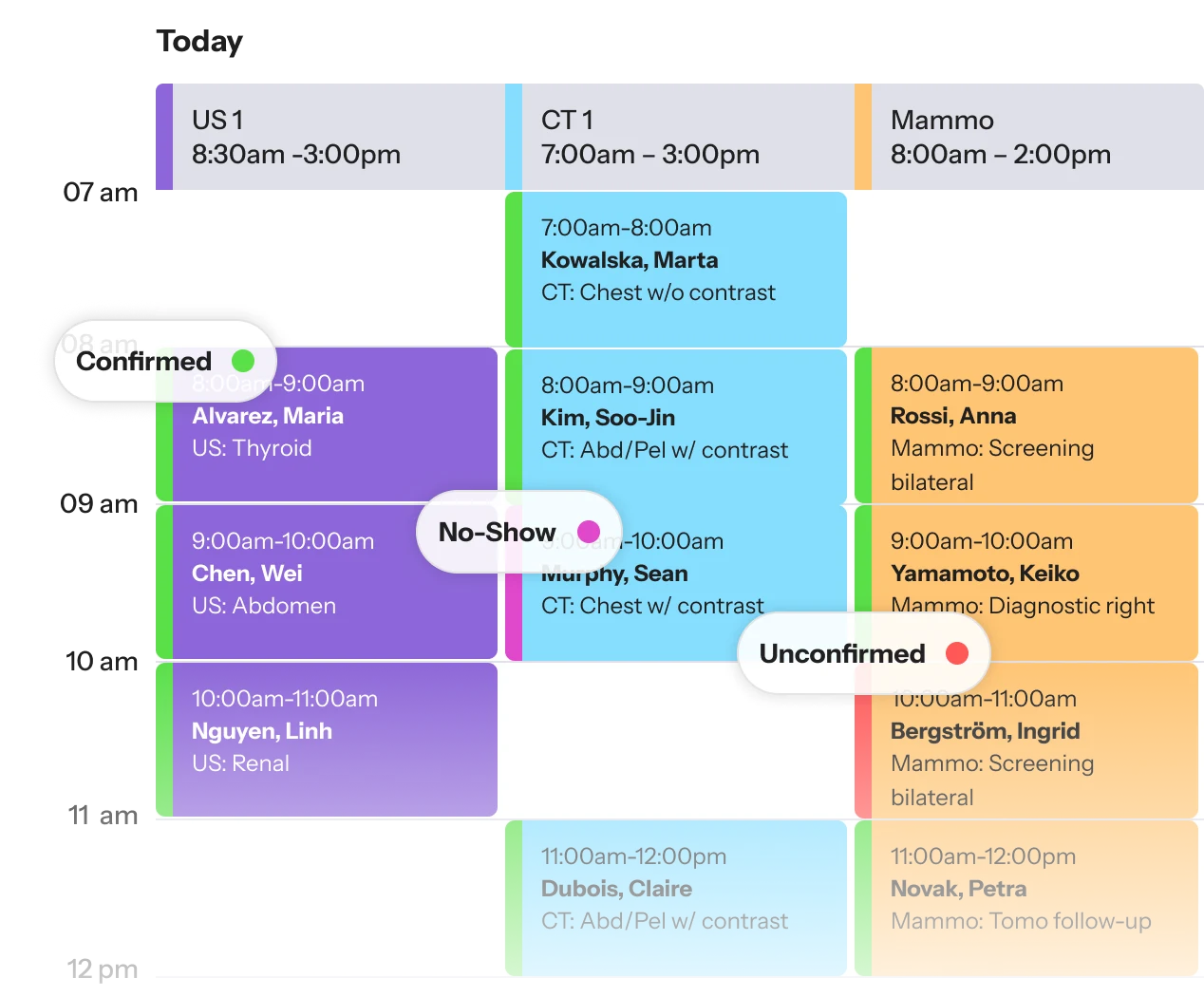
A full schedule depends on the right patient being ready for the right exam at the right time. Syneris brings scheduling, patient intake, orders and study status into one operational view.
- AI-powered scheduling
- Optimize scheduling intelligently around exam requirements, resources and appointment capacity while reducing administrative work.
- Online patient booking request
- Give patients the ability to request or book appointments online, extending access beyond the phone and front desk.
- Keep orders with the patient
- Keep the information needed to perform the study associated with the patient and exam as work moves from scheduling to acquisition.
- Know what’s happening now
- See what is scheduled, arrived, in progress, completed and ready for the next step without piecing the day together manually.
- See where capacity is going unused
- Coordinate appointment activity and available capacity across scanners and locations from a shared operational view.
PACS + diagnostic viewer
The study is complex. Getting to the read shouldn’t be.
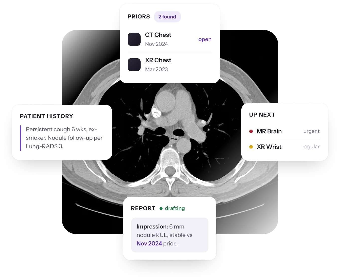
Imaging studies can contain hundreds or thousands of images across multiple series and priors. Syneris PACS brings the study, relevant imaging history and diagnostic tools into the reading environment, so radiologists can focus on interpretation.
- Current studies and relevant priors
- Access the imaging history needed for comparison without turning prior retrieval into a separate task.
- AI-assisted prior summary
- Surface key findings from relevant prior reports, helping radiologists understand the patient’s imaging history without reviewing each report individually.
- Modality-specific hanging protocols
- Present each modality’s studies in consistent viewing arrangements based on the way radiologists prefer to read.
- AI-powered report proofreader
- Uses AI to flag potential errors, inconsistencies and omissions before finalization, adding an extra layer of quality control.
- Multi-series navigation
- Move efficiently between sequences, series and image stacks while maintaining the context of the examination.
- Advanced visualization tools
- Use diagnostic viewing capabilities such as MPR, windowing, measurements and annotations within the PACS environment.
Billing
Protect the revenue behind every completed study.
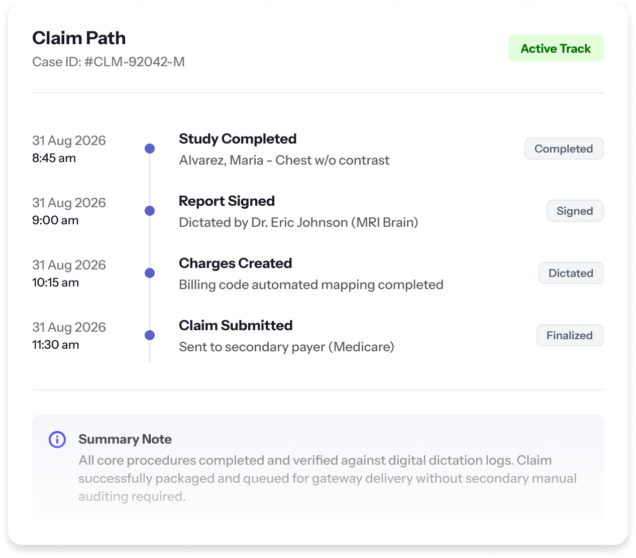
Billing problems often start upstream. Missing patient information, incomplete orders and inconsistent study details create work downstream. Syneris carries accurate information through the imaging process and into the billing and revenue-cycle systems your organization uses.
- Cleaner data from the start
- Carry patient, order and exam information forward so billing teams are not repeatedly reconstructing information created earlier in the imaging process.
- Billing and RCM connectivity
- Connect imaging operations with the billing platforms, clearinghouses and revenue-cycle partners your organization already uses.
- CPT-based clinic configuration
- Build your clinic database around the CPT codes you use, connecting procedures to the appropriate exam and billing information from the start.
- Documentation continuity
- Keep patient, order and study information with the exam to support accurate downstream documentation.
- Operational visibility before billing
- Identify incomplete or unresolved studies earlier in the process instead of discovering problems after they reach the revenue cycle.
Business intelligence & analytics
Turn imaging activity into operating intelligence.
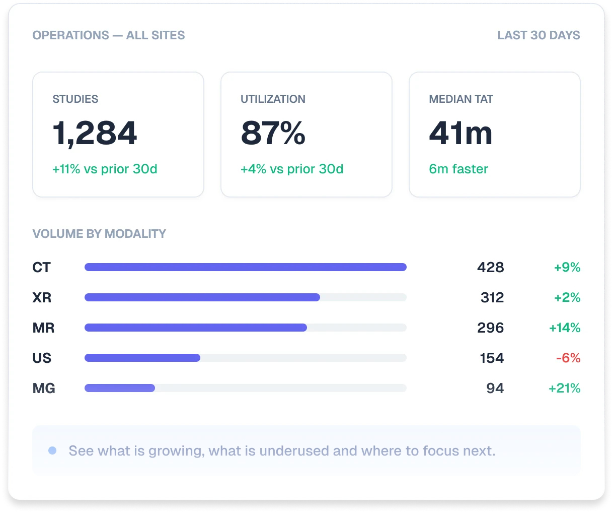
Volume alone doesn’t tell the whole story. Syneris shows how scanner utilization, appointment capacity, turnaround and performance vary across your operation, giving managers a clearer picture of where to act.
- Scanner capacity and utilization
- Track utilization across scanners, sites and time periods to identify gaps and make better use of capacity.
- Operational throughput
- Follow how studies progress through the imaging operation and identify where delays accumulate between scheduling, acquisition, interpretation and completion.
- Turnaround performance
- Monitor reporting turnaround and identify trends across modalities and locations.
- Scheduling performance
- Evaluate appointment activity, open capacity and scheduling patterns to understand where scanner time is being used effectively and where opportunity remains.
- Cross-site performance
- Compare operational activity across locations instead of managing each imaging center as an isolated view of the business.
Patient engagement & referring physician portal
Extend access beyond the imaging center.
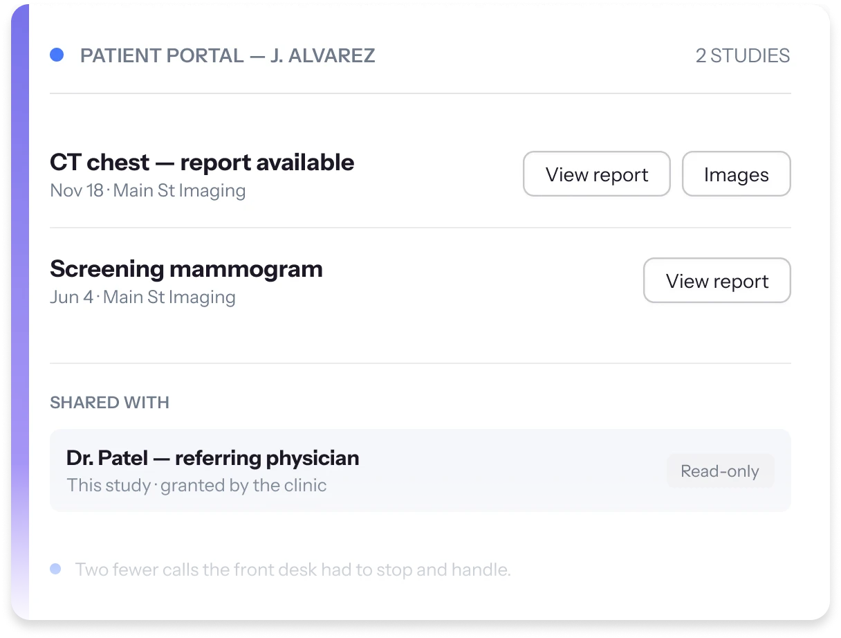
The exam may be complete, but the information still needs to move. Syneris gives patients and authorized referring physicians direct digital access to available images and reports, reducing manual follow-up for staff.
- Patient access to reports and images
- Give patients secure online access to available imaging information without making every request a phone call to the clinic.
- Referring physician access
- Allow authorized referring providers to retrieve patient reports and imaging through a dedicated portal.
- Browser-based image viewing
- Enable appropriate imaging to be reviewed online without requiring every external user to operate a diagnostic workstation.
- Faster report availability
- Make finalized information accessible to authorized users as it becomes available, helping reduce unnecessary administrative follow-up.
- Branded digital experience
- Extend the imaging center’s identity into the patient and referrer experience rather than sending users into a disconnected third-party environment.
Modality-specific tools
Tools shaped around the exam in front of you.
General imaging covers very different kinds of reading. These are the tools Syneris brings to each one.
- Hanging protocols
- Automatically arrange medical images and documents using predefined layouts to streamline review workflows and reduce manual setup.
- Window leveling and brightness/contrast
- Fine-tune image clarity to emphasize subtle anatomical differences and improve diagnostic accuracy.
- Annotation and measurement tools
- Mark up images and capture precise measurements for distance, area and volume.
- Split screen and split series
- View and compare multiple image series side by side, ideal for reviewing DICOM sets.
- Volume measurements
- Essential in obstetrics and gynecology for evaluating fetal and organ development.
- Structured reporting
- Generate reports that combine measurements, annotations and images directly from your ultrasound workflow.
- Cineloop autoplay and auto repeat
- Automatically play video clips to analyze movement and function, such as fetal activity or cardiac motion.
- Region of interest (ROI) analysis
- Evaluate specific skeletal regions like lumbar spine or hip for bone density metrics.
- Hanging protocols
- Streamline viewing by auto-applying standard display layouts for BMD studies.
- Annotation and measurement tools
- Use visual tools to assess and document measurements efficiently.
- Validation of patient eligibility for BMD
- Automatically confirm patient eligibility for baseline or follow-up BMD exams.
- Window leveling and brightness/contrast
- Adjust image tone and contrast to enhance visibility of tissues and bone structures.
- Magnification and zoom
- Focus on specific areas for more detailed examination.
- Measurement tools
- Assess dimensions, angles and regions within the X-ray image.
- Annotation and markup tools
- Add visual indicators like arrows, labels and shapes to guide clinical review.
- Image comparison
- Display current and past studies side by side to track progress or changes over time.
- Angle tools
- Measure spinal curvatures using Cobb angle and other precision tools.
Bring a study. We will walk through it live.
The fastest way to judge an imaging platform is to watch it handle the work you actually do all day.